Home » Without Label » Back Of Neck Anatomy / Anatomy Illustration Of Back Muscle Groups Showing Neck Pain 3d Illustration Stock Photo Alamy / Dummies helps everyone be more knowledgeable and confident in applying what they know.
Back Of Neck Anatomy / Anatomy Illustration Of Back Muscle Groups Showing Neck Pain 3d Illustration Stock Photo Alamy / Dummies helps everyone be more knowledgeable and confident in applying what they know.
Back Of Neck Anatomy / Anatomy Illustration Of Back Muscle Groups Showing Neck Pain 3d Illustration Stock Photo Alamy / Dummies helps everyone be more knowledgeable and confident in applying what they know.. It is made up of bones discs muscles ligaments nerves and tendons. Understanding the anatomy of your cervical spine and the vital nerves it contains should motivate you to adopt behaviors that help prevent neck injury and. In fact, the smallest muscle of the skeleton is the stapedius, which measures around 1 millimeter (1/20th of an inch) in length. « back show on map ». Levator scapulae, rhomboids (two), serratus posterior superior, splenius.
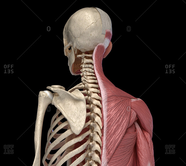
Head and neck trunk and limbs. Human anatomy diagrams and charts show internal organs, body systems, cells, conditions, sickness and symptoms information and/or tips to ensure one lives in good health. Beneath the integument the back of neck presents in the median plane the ligamentum nuchae, which is a triangular fibrous sheet and represents upward continuation of supraspinous ligament. The levator scapulae muscle is attached at the top four cervical vertebrae (c1 to c4) and runs down the side of the neck to attach at the top of the shoulder blade (scapula). In order to fully understand primary neck cancers, it helps to understand the anatomy and function of the structures in the neck.
3d Theeth Stock Photos Offset from ak.picdn.net Cervical fascia and interfascial spaces in the neck. « back show on map ». Levator scapulae, rhomboids (two), serratus posterior superior, splenius. Anterior muscles of the neck. Dummies helps everyone be more knowledgeable and confident in applying what they know. D) demonstrate sound knowledge of the surface/living and radiological anatomy. Posterior border of the ligament is free, anterior border is attached to the cervical spines and its superior border. The spine runs from the base of your skull down the length of your back, going all the way down to your pelvis.
When most people mention their back, what they are actually referring to is their spine.
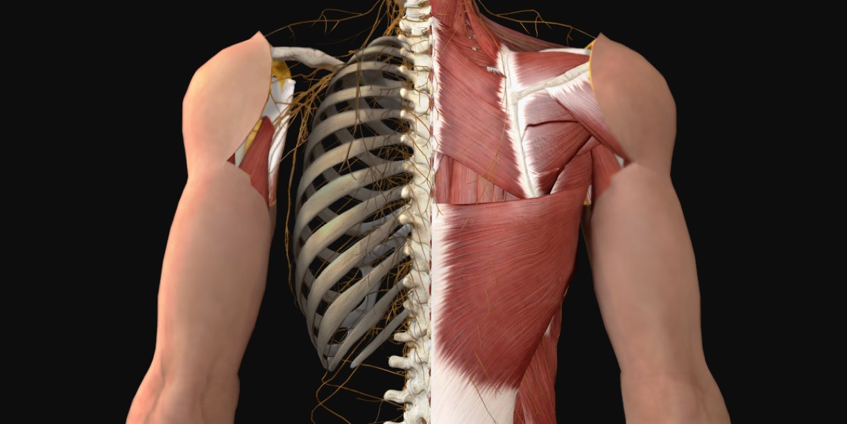
Digastric, mylohyoid, geniohyoid, stylohyoid infrahyoid muscles: Even the middle ear takes part in the muscular system of the head and neck. The splenius muscles originate at the midline and run laterally and superiorly to their insertions. He hath borne me on his back a thousand times. The spine runs from the base of your skull down the length of your back, going all the way down to your pelvis. Muscles of the posterior neck and the back. Your neck is like no other part of the vertebral spinal column and enables your head and neck a wide range of motion. The neck is a complex anatomic region between the head and the body. The levator scapulae muscle is attached at the top four cervical vertebrae (c1 to c4) and runs down the side of the neck to attach at the top of the shoulder blade (scapula). Posterior border of the ligament is free, anterior border is attached to the cervical spines and its superior border. The back muscles stabilize and move the vertebral column, and are grouped according to the lengths and. Whether it's to pass that big test, qualify for that big promotion or even master that cooking technique; E) demonstrate practical lab skills in anatomy and an appreciation of the ethics.
Posterior triangle of the neck boundari… pretracheal fascia b. Some important structures contained in or passing through the neck include the seven cervical vertebrae and enclosed spinal cord, the jugular veins and carotid arteries, part of the esophagus, the larynx. The physicians originally studying human anatomy thought the skull looked like an apple. Whether it's to pass that big test, qualify for that big promotion or even master that cooking technique; Clinically, surface anatomy is used to split the neck into anterior and posterior triangles which provide clues as to the location of specific structures.
Upper Cervical Spine Disorders Anatomy Of The Head And Upper Neck from www.spineuniverse.com Anatomic basis for local anesthesia. Levator scapulae, rhomboids (two), serratus posterior superior, splenius. The neck contains a number of overlapping muscles, blood vessels, nerves and myriad structures all contained in a small space and liable to damage and distress. Textbook of head and neck professor of anatomy (ret.) department of biomedical sciences baltimore college of dental. Dummies has always stood for taking on complex concepts and making them easy to understand. When to have lower back surgery. Clinically, surface anatomy is used to split the neck into anterior and posterior triangles which provide clues as to the location of specific structures. Posterior triangle of the neck boundari… pretracheal fascia b.
The cervical spine supports the weight and movement of your head and pro. We use cookies to ensure that we give you the best ex. The levator scapulae muscle is attached at the top four cervical vertebrae (c1 to c4) and runs down the side of the neck to attach at the top of the shoulder blade (scapula). Despite being a relatively small region, it contains a range of important anatomical features. Anatomy of male neck pain. Lymphatics of the head and neck. Muscle head anatomy vocal organ diagram female neck anatomy neck wireframe head neck human anatomy head artery anatomy face pharynx vector neck degree head anatomy 3d. The splenius muscles originate at the midline and run laterally and superiorly to their insertions. The neck is the area between the skull base and the clavicles. Of working with human remains; Submandibular triangle carotid and muscular triangles sternocleidomastoid region. He hath borne me on his back a thousand times. Your neck is like no other part of the vertebral spinal column and enables your head and neck a wide range of motion.
Cervical fascia and interfascial spaces in the neck. An overview of the anatomy of the hand, including the bones of the hand, muscles, blood supply and nerve supply. The cervical spine has seven vertebra of which the bottom five are designed similarly and the top 2 are very different. Muscles of the posterior neck and the back. Posterior triangle of the neck boundari… pretracheal fascia b.
Muscles Of The Back Anatomy Snippets Complete Anatomy from cdn.completeanatomy.cn He hath borne me on his back a thousand times. When most people mention their back, what they are actually referring to is their spine. Despite being a relatively small region, it contains a range of important anatomical features. Neck, in land vertebrates, the portion of the body joining the head to the shoulders and chest. Clinically, surface anatomy is used to split the neck into anterior and posterior triangles which provide clues as to the location of specific structures. This human anatomy diagram with labels depicts and explains the details and or parts of the back of neck anatomy. Muscles of the posterior neck and the back. The neck or cervical spine is the top part of the spine between the head and shoulders.
In fact, the smallest muscle of the skeleton is the stapedius, which measures around 1 millimeter (1/20th of an inch) in length.
This article describes the anatomy of the head and neck of the human body, including the brain, bones, muscles, blood vessels, nerves, glands, nose, mouth, teeth, tongue, and throat. Your neck is like no other part of the vertebral spinal column and enables your head and neck a wide range of motion. It is made up of bones discs muscles ligaments nerves and tendons. When to have lower back surgery. In order to fully understand primary neck cancers, it helps to understand the anatomy and function of the structures in the neck. Anatomy of male neck pain. Neck muscles help support the cervical spine and contribute to movements of the head, neck, upper back, and shoulders. Digastric, mylohyoid, geniohyoid, stylohyoid infrahyoid muscles: Neck, in land vertebrates, the portion of the body joining the head to the shoulders and chest. The neck is the area between the skull base and the clavicles. The cervical spine supports the weight and movement of your head and pro. Whether it's to pass that big test, qualify for that big promotion or even master that cooking technique; Clinically, surface anatomy is used to split the neck into anterior and posterior triangles which provide clues as to the location of specific structures.